
| OVERVIEW | HIGHLIGHTS | PROJECTS | PEOPLE |
 One treatment for brain tumors in the head is to cut off the tumor's
blood supply--its source of nourishment--and, thus, destroy the
tumor. This is accomplished by introducing a catheter in the groin and
snaking it into the blood vessels that supply the tumor. Emboli are
then injected to block that blood supply. The positioning of the
catheter is presently done under the guidance of X-ray angiography,
with all route planning done in the operating room. The X-ray dose
and burden of toxic opacifying material is extraordinarily high.
Also, because the X-ray angiograms are 2D projections, difficulty in
understanding the 3D course of the vessels leads to false steps that
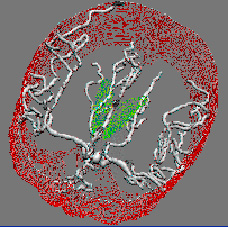
greatly increase the time of surgery. In the method we are
developing, 3D imaging of the intracranial blood vessels by magnetic
resonance
or CT angiography and multiplane (i.e., multipose) X-ray angiography
are done prior to surgery, and these images are analyzed to produce a
3D map of the blood vessel area in which we are interested. The
catheter route is planned before surgery using computer graphics and
this 3D map. During surgery, the relevant portion of the 3D vascular
tree in which the catheter lies will be shown from approximately the
same pose as each intraoperatively acquired X-ray angiogram, allowing
the clinician to interpret each 2D image within a 3D context. With
this technique, we expect to see a significant decrease in the amount
of patient X-ray exposure, a decrease in the length of the surgery,
and an increase in the accuracy in positioning the catheter.
One treatment for brain tumors in the head is to cut off the tumor's
blood supply--its source of nourishment--and, thus, destroy the
tumor. This is accomplished by introducing a catheter in the groin and
snaking it into the blood vessels that supply the tumor. Emboli are
then injected to block that blood supply. The positioning of the
catheter is presently done under the guidance of X-ray angiography,
with all route planning done in the operating room. The X-ray dose
and burden of toxic opacifying material is extraordinarily high.
Also, because the X-ray angiograms are 2D projections, difficulty in
understanding the 3D course of the vessels leads to false steps that
greatly increase the time of surgery. In the method we are
developing, 3D imaging of the intracranial blood vessels by magnetic
resonance
or CT angiography and multiplane (i.e., multipose) X-ray angiography
are done prior to surgery, and these images are analyzed to produce a
3D map of the blood vessel area in which we are interested. The
catheter route is planned before surgery using computer graphics and
this 3D map. During surgery, the relevant portion of the 3D vascular
tree in which the catheter lies will be shown from approximately the
same pose as each intraoperatively acquired X-ray angiogram, allowing
the clinician to interpret each 2D image within a 3D context. With
this technique, we expect to see a significant decrease in the amount
of patient X-ray exposure, a decrease in the length of the surgery,
and an increase in the accuracy in positioning the catheter.To achieve these aims, intensive computational resources are needed, particularly for the 3D vessel reconstruction and real-time registration of CT/MRI with X-ray images. Before receiving the grant from the Intel Technology for Education 2000 Program, this work was being done on high-end specialty computers. The Intel gift has made possible a switch to the use of computers based on standard Intel and Windows NT components, and this configuration can be easily reproduced in the many other hospitals in the U.S. and around the world where this surgical procedure takes place.
For additional information about this research, see the projects page on the Computer Aided Diagnosis and Display Lab web page.