UNC Laparoscopic Visualization Research

Structured Light Experiments
on a Pig
utilizing DMD projector, laparoscope and Flexscope


  These images were taken from experiments performed at the University of North Carolina medical school on an ex-pig.  Experiments were performed by the computer science department to determine the problems associated with 3D image extraction by structured light in a body cavity.   These images then are reconstruceted in an HMD to assist in minimally invasive surgical procedures.   Some more details of this work can be found on the UNC computer science webpages for the laparoscopic visualizatiuon research  and 3D ultrasound research.  This research and patents for the technology have been licensed by InnerOptic Technology, Inc. and are presently commercializing this system
  These experiments were conducted  June 24th 1998  by:

Michael Rosenthal
Jeremy Ackerman
Kurtis Keller

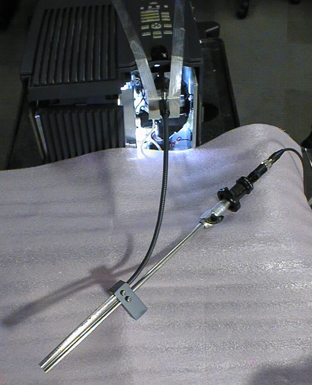
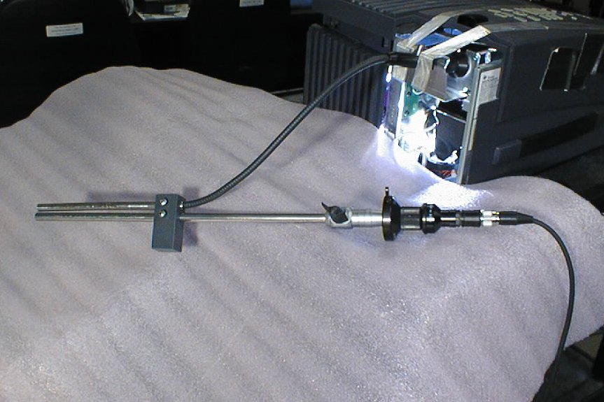
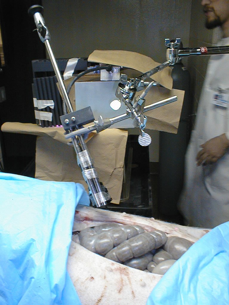
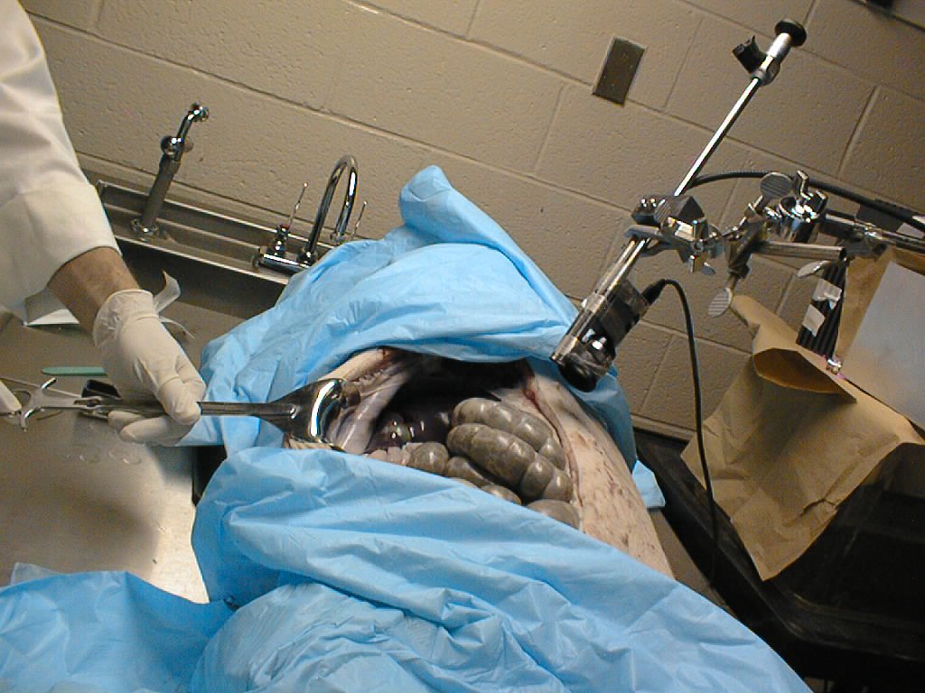
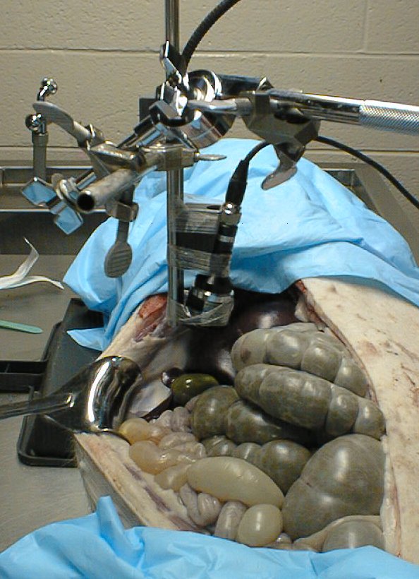
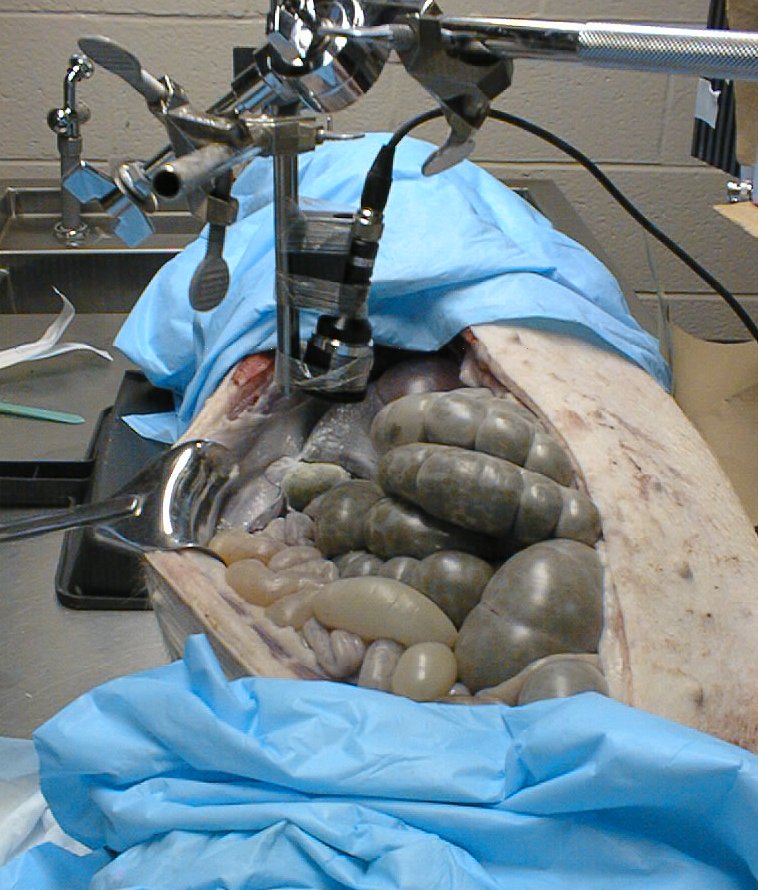
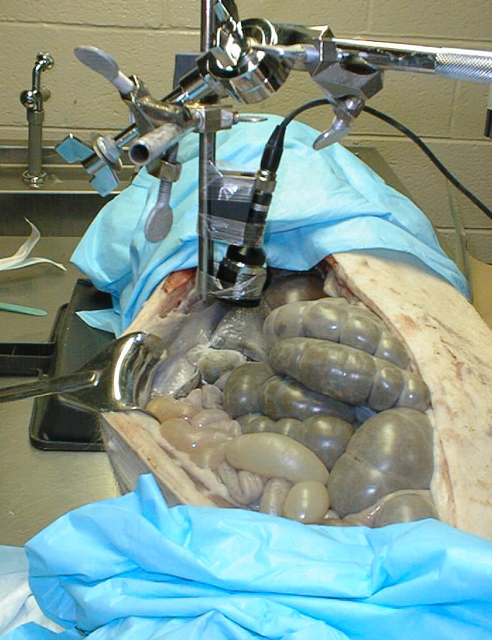
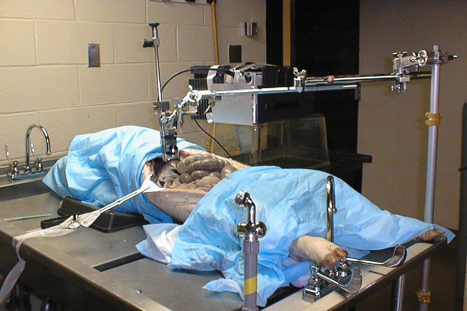
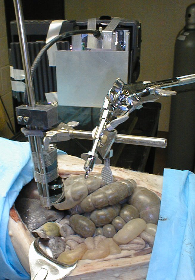
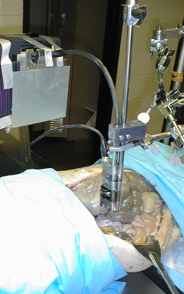
  Here are two pictures of the laparascope / fiberscope setup:

Flex scope hooked up to DMD deviceFlex scope hooked up to DMD device

  Here is the next generation laparascope / structured light projector in its actual, miniaturized form.  This  item is only viewable inside the computer science domain.
                        New Projector / Laparascope animated gif

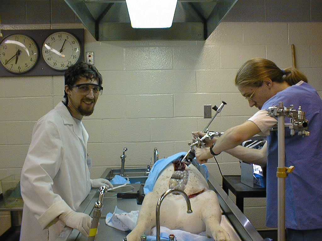
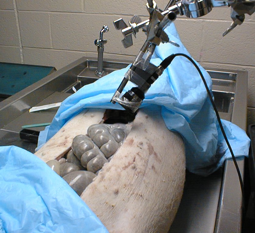
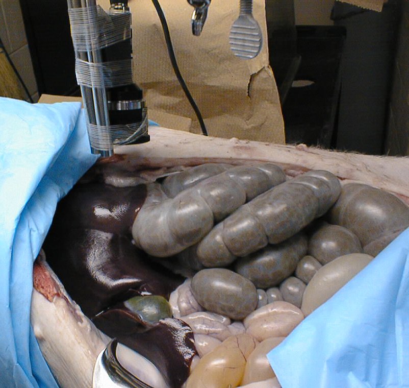
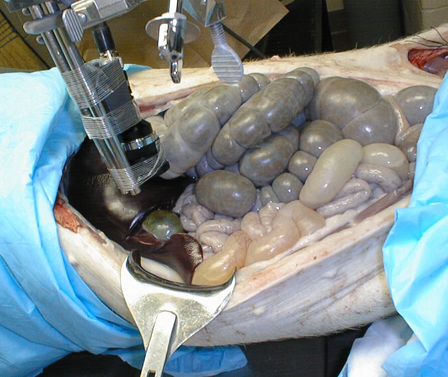
  Following are pictures from the actual experiments.  The thumbnails are small to avoid showing much detail because some of them are rather gross.  These are very graphic pictures of a pig opened up.  You are warned:

Michael and Jeremy with the pigpig with laparascopepig with laparascope


pig with laparascopelaparascope aimed at gall bladderlaparascope aimed at gall bladder
 

laparascope aimed at gall bladderlaparascope aimed at gall bladderwith talc powder
 

Identity of pig concealed


All images Copyright 1998 UNC.