UNC Ultrasound/Medical Augmented Reality Research
Augmented Reality Technology
Overview
Our research group is working to develop and operate a system that allows
a physician to see directly inside a patient, using augmented reality (AR).
AR combines computer graphics with images of the real world. This project
uses ultrasound echography imaging, laparoscopic range imaging, a video
see-through head-mounted display (HMD), and a high-performance graphics
computer to create live images that combine computer-generated imagery
with the live video image of a patient. An AR system displaying live ultrasound
data or laparoscopic range data in real time and properly registered to
the part of the patient that is being scanned could be a powerful and intuitive
tool that could be used to assist and to guide the physician during various
types of ultrasound-guided and laparoscopic procedures.
Head Mounted Display Research
Head mounted displays (HMDs) with appropriate characteristics for augmented
reality are not commercially available. We have put a significant effort
into modifying existing HMDs and building
our own HMDs for use in our augmented reality projects.
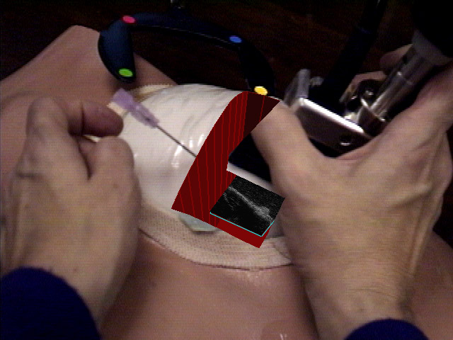
Ultrasound-guided Breast Biopsy
In recent years, ultrasound-guided biopsy of breast lesions has been
used for diagnostic purposes, partially replacing open surgical intervention.
Ultrasound guidance is also often used for needle localization of some
lesions prior to biopsy, as well as for cyst aspiration. Ultrasound guidance
for such interventions, however, is difficult to learn and perform. One
needs good hand-eye coordination and three-dimensional visualization skills
to guide the biopsy needle to the target tissue area with the aid of ultrasound
imagery. We believe that the use of computer-augmented vision technology
can significantly simplify both learning and performing ultrasound-guided
interventions. We are, therefore, targeting our current and near-term future
research efforts toward building a system that will aid a physician in
performing an ultrasound-guided needle biopsy. Results from preliminary
experiments with phantoms and with one human subject have been encouraging.
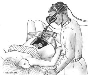
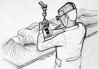
Early Augmented-Reality Systems.
Our initial ultrasound visualization system, featured the ability to display
a small number of individual ultrasound slices of a fetus superimposed
onto the pregnant patient's abdomen. The system used traditional chroma-keying
techniques to combine rendered ultrasound data with a digitized video image
from a head-mounted camera. In the initial system, alignment was poor (due
to poor tracking), the images were not clear, and the sense of the 3D shape
of the fetus was lacking. The first improvement to the system was to reconstruct
the individual slices of ultrasound data into a volume. This algorithm
made use of UNC's custom high-performance graphics engine, Pixel-Planes
5. Updates were slow, however, and the images were still not convincing.
We thus moved to a system that featured on-line data acquisition and off-line
rendering. These images set a quality standard for later systems, though
we still wanted to return to a real-time system. Also, as the supporting
technologies improved, we incorporated these into the system. Alignment
was improved by new camera and ultrasound probe calibration methods, and
by improvements in instrument and head tracking.
Intermediate Real-Time System.
The ultrasound research group moved to a prototype real-time augmented
reality system based on an SGI Onyx Infinite Reality (IR) high-performance
graphics workstation equipped with a Sirius Video real-time capture unit.
This system made use of the high-speed, image-based texturing capability
available in the IR. The Sirius captured both HMD camera video and ultrasound
video. The camera video was displayed in the background; the ultrasound
video images are transferred into texture memory and displayed on polygons
emitted by the ultrasound probe inside a synthetic opening within the scanned
patient. The display this system presented to the user resembles the display
offered by the earlier on-line volume reconstruction system, but the images
obtained are vastly superior to those generated by the initial system.
This system could sustain a frame rate of 10 Hz for both display update
and ultrasound video grabbing and also provides high-resolution ultrasound
slice display and rendering for up to 16 million ultrasound pixels. This
system used a technique designed to correct image errors from the
magnetic head tracking system by tracking landmarks in the video imagery.
Other techniques such as prediction, interpolation of past readings, and
reordering of computation in order to reduce apparent latency have proven
useful in further reducing registration errors.
Unlike the initial system, the ultrasound slices are not reconstructed
into a volume, but are displayed directly as translucent polygons with
ultrasound video images mapped on them. A large number of such directly
rendered ultrasound slices can give the appearance of a volume data set.
Ultrasound probe tracking was performed with a high-precision, mechanical
tracking device that provided correct registration between individual ultrasound
slices.
This system was used to demonstrate the possibility of using augmented
reality to enhance visualization for laparoscopic
surgery .
Current Real-Time System
We recently moved our prototype real-time augmented reality system from
an SGI Onyx InfiniteReality(tm) to the departments SGI Reality Monster.
We make use of the digital video input capabilities of the Reality Monster
by simultaneously capturing imagery from the HMD cameras, and the ultrasound
imager into texture memory. The new system uses only a single opto-electric
tracking system with greater overall precision than previous systems.
New applications of our system require advances in tracking, image processing,
and display devices. In collaboration with the University of Utah, we designed
and built a video see-through head-mounted display and are experimenting
with alternatives to HMDs. Currently available tracking systems limit our
ability to precisely register real and synthetic imagery leading to research
into improving tracking systems. As we began to develop our system for
laparoscopic surgery, we have found a need to develop instruments and algorithms
for rapidly acquiring the three dimensional geometry inside a body through
a laparoscope.
Past Subprojects for Augmented Reality
In building this system, we have worked extensively on (what is known in
the literature as) augmented reality technology, especially on registration
of the real and synthetic worlds, both spatially and temporally. There
are three areas that we have invested a significant research effort in
the past few years.. Each is described on a separate web page.
-
Hybrid tracking
Our systems used to achieve registration of the real and virtual worlds
by integrating vision-based and magnetic trackers.
-
Magnetic tracker calibration
Calibration of the magnetic tracker aids the hybrid tracking algorithm
above, but cannot alone yield adequate registration. In fact, it turns
out to be very difficult to do and rather impractical.
-
Latency management
Realistic integration of the virtual world with the dynamic real world
creates additional problem to be solved..
Research Sponsors
Last Modified: June 15, 2000 by Jeremy
Ackerman
For more info send email to Andrei State at andrei[at-sign]cs.unc.edu