
MIDAG Celebrates 40 Years
The MIDAG 40th Anniversary Celebration was held at the Carolina Inn and in Sitterson Hall on October 18th and 19th. For a recap of the events, please click here.
The Medical Image Display and Analysis Group (MIDAG) will celebrate its 40th anniversary this academic year concurrently with the department’s 50th anniversary.
How did a research group so dependent on collaboration between departments in different scientific disciplines come to be?
MIDAG’s roots can be traced all the way back to Massachusetts General Hospital in the mid-1960s, where Harvard University doctoral student Steve Pizer was working on the world’s first dissertation in medical imaging. His work on scintigraphic image restoration for diagnostic tests in nuclear medicine led to an invitation to present at the Second International Conference on Information Processing in Medical Imaging.
Even though IPMI 2 never came to be, its planning stages resulted in an agreement between Pizer and Andrew Todd-Pokropek that Pizer would take a year-long sabbatical at University College Hospital in London. His work with Todd-Pokropek in London led to the realization that restoration improvements were being nullified by inadequate approaches to medical image display. It was then that Pizer decided to re-focus his research efforts onto medical image display. When he returned to UNC in 1974, he sought out interdepartmental collaboration to form what is now MIDAG.
With Gene Johnston and Ed Staab having recently joined the UNC Radiology Department from Vanderbilt, Pizer found two UNC faculty members with whom collaboration would afford unique opportunities for multidisciplinary advancements.
Early collaborations focused on two-dimensional medical image display, which continued into the 21st Century. Adaptive contrast enhancement research proved, for perhaps the first time, that increased care in the mapping of intensity between the recorded and displayed results had a significant positive impact on the accuracy of diagnostic or therapeutic decisions. This approach to research by first examining the techniques became a cornerstone of the MIDAG philosophy.
MIDAG research into standardization of electronic displays led to the adoption of international ACR-NEMA standards for transmission of medical images, which greatly improved the ability of radiologists and medical physicists to decode output images.
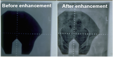
This pelvic portal image provides an example of the
benefits of contrast enhancement techniques
Another important area of two-dimensional MIDAG research was human vision. Visual psychophysics, the study of the relationship between visual stimuli and physiological sensation and perception, provided a groundwork for better understanding how medical images are interpreted.
When Henry Fuchs joined the Department of Computer Science in 1978, an additional focus of MIDAG research emerged: three-dimensional display.
Using the varifocal mirror, Fuchs, Pizer, and Duke neuroradiologist Ralph Heinz determined that three-dimensional visualization had merit, but not without occlusion, or the hiding of elements that shouldn’t be visible in order to de-clutter the image.
Important contributions from MIDAG doctoral student Marc Levoy led to the invention of new methods of rendering through ray casting, as well as the application of those methods in radiation treatment planning under the leadership of UNC Radiation Oncology’s Julian Rosenman.
Pizer likes to recall that ray casting and splatting, two methods of three-dimensional rendering, were both invented by members of the UNC Computer Science team (Marc Levoy and Lee Westover, respectively) and that MIDAG was among the first research groups to undertake work in the field of three-dimensional rendering.
Image analysis would become a very large part of MIDAG’s research in the 1980s. MIDAG research in the area of object representation using medial geometry contributed to the representation of anatomic objects in the real world medially via the “m-reps” structure.
Fast-forward to the 2014-2015 academic year, when the UNC Medical Image Display and Analysis Group will be celebrating its 40th Anniversary. Its current membership includes the Departments of Computer Science, Biomedical Engineering, Radiology, Surgery, Psychiatry, Radiation Oncology, Mathematics, Biostatistics, Statistics, and Neurology, as well as the Schools of Dentistry and Information & Library Science. Considering the relative youth of many of the disciplines involved in MIDAG research further underlines the impressiveness of both the longevity and the significant level of contribution that MIDAG has made over the past 40 years.
Embracing the philosophies set down by department heads such as Frederick P. Brooks, Jr. (Computer Science) and Ed Chaney (Radiation Oncology), the research group has flourished and continued to grow and contribute to medical advances despite the notoriously long time required to take a medical breakthrough from concept to implementation.